
Pupil Dilation Explained: What to Expect at Your Eye Exam
Pupil dilation typically causes 4–6 hours of blurred near vision and light sensitivity, but it remains the gold standard for detecting sight-threatening diseases like glaucoma and retinal tears. At Frame & Focus Eye Care, Dr. Sarah Zaver uses this essential 30-minute procedure to gain a comprehensive 3D view of your retina and optic nerve that digital imaging simply cannot match.
Schedule Your Comprehensive Eye Exam in Richmond
Why Dilation Is the Gold Standard for Eye Health
When you look into someone’s eye, the pupil—the black center—acts like a window. Normally, the pupil constricts (gets smaller) when exposed to bright light, which limits your doctor’s view to only the central part of the eye. Think of it like looking through a keyhole into a dark room.
Pupil dilation temporarily forces this window wide open, giving your doctor a full, panoramic view of the internal structures. This comprehensive view is why the American Academy of Ophthalmology (AAO) and the National Eye Institute (NEI) strongly recommend regular dilated exams. The procedure transforms the inspection from a limited central view to a detailed, three-dimensional assessment of the entire posterior segment of the eye.
What Your Doctor Sees: The Crucial Internal Structures
Dilation allows your optometrist to examine three non-negotiable areas critical for long-term vision health:
-
The Retina: This light-sensitive layer at the very back of the eye functions like the film in a camera. Dilation allows the doctor to check for tears, holes, swelling, or thinning—all signs of diseases that have no outward symptoms until significant vision loss has already occurred.
-
The Optic Nerve: This is the ‘cable’ that transmits all visual information from the retina to the brain. The optic nerve head has a specific appearance, and any changes in its cup-to-disc ratio or color can be the very first sign of glaucoma—a disease often called the “silent thief of sight.”
-
The Macula: The macula is the small central portion of the retina responsible for sharp, detailed central vision (the vision you use for reading or recognizing faces). Checking the macula is essential for detecting the early stages of Age-Related Macular Degeneration (AMD).
The Consequences of Skipping Dilation
Opting out of dilation in favor of non-dilated imaging, while convenient, carries a significant clinical risk. Without dilation, a doctor risks missing pathology in the far periphery, which can lead to delayed diagnosis and irreversible vision loss.
|
Missed Condition |
Long-Term Consequences of Delayed Detection |
|---|---|
|
Peripheral Retinal Tear/Hole |
Often asymptomatic until the tear progresses to a full Retinal Detachment, which is a vision-threatening emergency requiring immediate, complex surgery. |
|
Early Glaucoma Changes |
Progressive, irreversible damage to the optic nerve. Vision loss begins in the peripheral field, making it undetectable by the patient until advanced stages. |
|
Subtle Diabetic Retinopathy |
Early micro-aneurysms or fluid leakage may be confined to the far retina. Missing these delays in treatment (like laser therapy or injections) can lead to macular swelling and central vision loss. |
Crucial for Early Disease Detection
For many serious eye conditions, early detection is the only path to successful management and vision preservation. A dilated eye exam can identify problems even before you experience any noticeable symptoms.
|
Condition Detected |
Why Dilation is Necessary |
|---|---|
|
Diabetes damages the tiny blood vessels in the retina. Dilation lets the doctor see subtle leakage, swelling, or abnormal vessel growth (neovascularization) before it causes permanent damage. |
|
|
Glaucoma |
Glaucoma damages the optic nerve. Dilation allows the doctor to inspect the entire optic nerve head for structural changes that indicate elevated pressure or nerve damage. |
|
Macular Degeneration |
Dilation helps spot drusen (tiny yellow deposits) or pigment changes in the macula, which are early markers of AMD. |
|
Retinal Detachment/Tears |
These emergencies require a full view of the peripheral retina, which is only possible with complete pupil dilation. |
|
Systemic Health Indicators |
Your eyes’ blood vessels are the only ones in the body that can be directly viewed. Changes can indicate serious systemic issues like high blood pressure or atherosclerosis. |
The Science Behind Pupil Dilation: How the Drops Work
Understanding how the dilation process works can significantly reduce any worry you might have.
Mydriatics vs. Cycloplegics: The Pharmacology of Dilation
The drops used in a dilated eye exam are not just one chemical, but often a combination of two types of medication, administered by your eye doctor in precise amounts to achieve maximal widening (mydriasis) and to eliminate the eye’s natural light-response mechanism (cycloplegia).
-
Mydriatic Agents (The Wideners – e.g., Phenylephrine): These drops are sympathomimetic, meaning they stimulate the radial (dilator) muscles of the iris, causing the pupil to actively widen. This effect is relatively quick, often starting within 10-15 minutes.
-
Cycloplegic Agents (The Relaxers – e.g., Tropicamide): These drops are anti-muscarinic. They paralyze two specific muscles:
-
The iris sphincter muscle ensures the pupil cannot constrict, even when exposed to bright light.
-
The ciliary body muscle which is responsible for accommodation (focusing). Paralyzing this muscle temporarily prevents the eye from focusing up close, which is the primary cause of temporary near vision blur.
-
When administered, you may feel a mild, brief stinging sensation that typically lasts only a few seconds. This is completely normal and means the drops are starting to work!
Why Some Eyes Dilate Faster (The Eye Color Factor)
Have you ever wondered why the person next to you seems to recover faster? Eye color is a key factor in the duration of dilation due to the amount of melanin pigment present in the iris.
-
Lighter Eyes (Blue, Green, Hazel): Individuals with lighter-colored irises often experience longer-lasting dilation effects (sometimes up to 6-8 hours). This is because lighter eyes have less pigment (melanin) to absorb, bind, and metabolize the medication, leaving more drug available to act on the muscles.
-
Darker Eyes (Brown, Dark Brown): People with darker irises often metabolize the drops more quickly due to higher melanin levels, resulting in shorter dilation duration (typically 4–6 hours).
Safety First: Narrow Angle Screening and Contraindications
Before Dr. Zaver administers the drops, she performs a crucial safety step: checking for a condition called narrow angles. This condition, where the iris is positioned close to the drainage meshwork of the eye, poses a risk. In rare cases, dilating the pupils of a patient with narrow angles could rapidly increase the internal eye pressure, potentially triggering an acute angle-closure glaucoma attack.
This is a critical safety measure, and another example of the thoroughness Dr. Zaver and the Frame & Focus team prioritize. If you have narrow angles, your doctor will discuss alternative examination methods or use a different, reversible dilating agent if needed. We also screen for known allergies to ensure patient safety.
Your Dilated Eye Exam: A Step-by-Step Timeline
A dilated eye exam is an efficient and painless process when you know what to expect. While the total visit may take longer than a routine check-up, the actual examination time is brief.
1. Pre-Exam Preparation
Before your appointment at Frame & Focus, we recommend:
-
Arranging Transportation: Even if you feel comfortable driving after previous dilations, we strongly advise having a friend, family member, or rideshare service take you home. Your eyes will be highly sensitive to glare, and your near vision will be impaired.
-
Bring Sunglasses: While we always provide disposable wraparound shades, bringing your own high-quality, polarized sunglasses will ensure maximum comfort on your drive home.
-
Gather Information: Have your current list of medications (including eye drops) and insurance information ready.
2. The Dilation Phase (15–30 Minutes)
-
Drop Administration (0-5 minutes): The doctor or technician will administer the dilating drops to both eyes. You may feel a brief sting.
-
The Waiting Period (15-30 minutes): This is the time it takes for the drops to fully relax your iris muscles and widen your pupils. During this period, you may notice your vision getting slightly blurry, especially up close, and light sensitivity beginning to increase. This is a great time to listen to a podcast or an audiobook!
3. The Examination Phase (10–15 Minutes)
Once your pupils are wide open, Dr. Zaver will begin the detailed inspection.
-
Slit Lamp Biomicroscopy: The doctor uses a high-powered microscope (the slit lamp) and specialized high-plus lenses to look through your widened pupil at the internal structures of your eye in three dimensions. This 3D view is essential for assessing the depth and contour of the optic nerve head.
-
Indirect Ophthalmoscopy: The doctor uses a head-mounted light and a handheld lens to look at the far periphery of your retina, ensuring a 360-degree view. This is crucial for catching subtle, hidden retinal issues.
4. Immediate Post-Exam: First Hour
After the exam, the doctor will discuss their findings with you. At this point, the effects of the drops are usually at their peak:
-
Near Vision Blur: You will find it very difficult to read or focus on close objects.
-
Photophobia (Light Sensitivity): Direct sunlight and bright overhead lights will feel intense. This is why those sunglasses are so important!

Managing Post-Dilation Side Effects and Recovery
While pupil dilation side effects are temporary, managing them effectively is key to a comfortable experience.
Light Sensitivity and Blurred Vision: The Two Main Effects
The symptoms you feel are a direct result of the drops’ mechanism of action:
-
Extreme Light Sensitivity: Your pupil is responsible for restricting light entry. With the pupil artificially wide open, more light floods the retina. This is particularly noticeable in bright sunlight. Actionable Tip: Wear dark, wraparound sunglasses continuously if you are outside or in a brightly lit indoor space.
-
Blurred Near Vision: The cycloplegic drops paralyze the ciliary muscle, preventing your eye from accommodating (flexing the lens to focus up close). This means objects at arm’s length or closer will appear blurry. Your distance vision, however, is often less affected, or sometimes even slightly enhanced, due to the increased light.
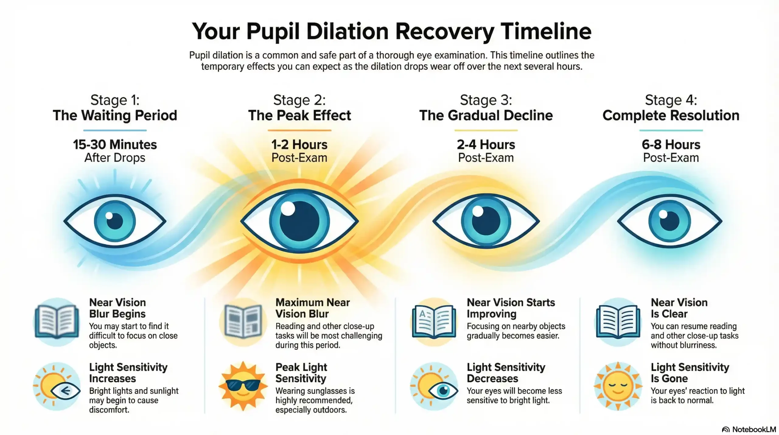
How Long Does Pupil Dilation Last? (Detailed Duration Table)
The question, “How long does eye dilation last?” is the most common one we hear. While the average is 4–6 hours, duration varies based on the type of drops used and your unique physiology (like your eye color and age).
|
Dilation Phase |
Typical Duration |
What to Expect |
|---|---|---|
|
Peak Effect |
1–2 hours post-exam |
Maximum blurriness and light sensitivity. Avoid screens and driving. |
|
Gradual Decline |
2–4 hours post-exam |
Near vision may begin to return, and sensitivity decreases slightly. Still not safe to drive. |
|
Near-Normal |
4–6 hours post-exam |
Most effects are gone for the majority of people. Near vision is functional, but night vision may still be slightly compromised. |
|
Complete Resolution |
6–8 hours (or up to 24 hours for children/light eyes) |
Pupils return entirely to normal size and reactivity. |
Essential Aftercare Tips for a Smooth Recovery
-
Plan for Screen-Free Time: Use the recovery period as an excuse to relax. Listen to music, an audiobook, or take a nap. If you must use a screen, increase the text size significantly and turn the screen brightness all the way down.
-
Stay Hydrated: This helps your body metabolize the drops.
-
Keep Your Hands Clean: Avoid rubbing your eyes, especially if they feel slightly irritated. This prevents introducing bacteria.
-
Try the Nearsighted Trick: If you are nearsighted (myopic), try removing your glasses completely. You may be able to read fine print at a very close distance because your focus is naturally tuned to near objects.
-
For Prolonged Effects: If dilation lasts longer than 8 hours, especially accompanied by a headache or severe eye pain, contact Frame & Focus Eye Care immediately. While rare, this could indicate an adverse reaction.
Driving and Daily Life: Planning for Your Post-Exam Hours
One of the most important aspects of preparing for your pupil dilation is managing your logistics afterward.
Can I Drive Myself Home? The Safety Verdict
While some patients feel comfortable driving themselves home, Frame & Focus Eye Care strongly advises against it, especially for first-time dilation. We prioritize your safety and the safety of the community in Richmond, Sugar Land, and Fulshear.
Here’s why driving is risky post-dilation:
-
Impaired Light Adaptation: Driving exposes you to unexpected glare from oncoming traffic, reflections, and sudden bursts of sun. Because your pupils cannot constrict, this glare is blinding and dangerous, reducing reaction time.
-
Lack of Near Focus for Dash/GPS: You need functional near vision to read your dashboard, car controls, or GPS screen. Since your near vision is temporarily blurred by the cycloplegic agents, operating a vehicle is compromised.
Please arrange a ride or plan to take a taxi/rideshare service. This small planning step prevents a potentially serious risk.
Using Digital Devices (Phone and Computer) During Recovery
If your job or daily routine requires screen time, plan to handle light tasks that do not require fine detail for the first few hours.
-
Maximize Accessibility Settings: On your phone or computer, go into the accessibility settings and increase font and display sizes to their maximum.
-
Use Dark Mode: Switching to dark themes on all apps and browsers can dramatically reduce the amount of bright light hitting your eyes.
-
Use Voice Commands: Navigate your phone and send texts using voice assistance to avoid straining your blurry near vision.
-
Consider Print: If you absolutely must read a document, print it out in a large, bold font. The contrast of black text on a white background is often more readable than a backlit screen when your pupils are dilated.
Special Consideration: Pupil Dilation for Children (Pediatric Patients)
The dilation process for children and teenagers is often slightly different but equally essential, especially for services like myopia management.
Why Pediatric Dilation is Critical
In children, dilation is performed not just to check the retina, but also to obtain the most accurate measure of their refractive error (their glasses prescription).
-
Pseudomyopia Prevention: Children’s eye muscles are powerful and can over-focus, which can mask hyperopia (farsightedness) or make myopia (nearsightedness) appear worse than it is. This is known as pseudomyopia or a latent hyperopia.
-
Cycloplegic Refraction: By paralyzing the focusing muscle with stronger cycloplegic drops (often a different formula than for adults, like Atropine or Cyclopentolate), Dr. Zaver can obtain a true, objective measurement of the child’s prescription, ensuring they get the correct glasses and myopia control treatments.
Pediatric Dilation Duration
Because children’s ciliary muscles are highly active, the cycloplegic drops used are often stronger and designed to last longer—sometimes up to 24 hours. The Frame & Focus team will discuss the exact timeline and aftercare to ensure your child remains comfortable and safe.
Dilation Alternatives: Digital Imaging vs. The Gold Standard
You may have heard about alternatives like Optomap or Digital Retinal Imaging, which are often offered as ways to avoid dilation. These technologies are powerful and utilize high-resolution cameras to capture detailed images of the retina without drops.
The Role of Retinal Imaging (Optomap)
At Frame & Focus Eye Care, we embrace state-of-the-art technology, including high-definition retinal imaging.
-
The Benefit: Imaging is quick, comfortable, provides a permanent digital record for year-to-year comparison, and captures up to 80% of the retina. This is an excellent screening tool for low-risk, routine follow-up patients.
-
The Limitation: While excellent for the central and mid-peripheral retina, digital imaging often cannot capture the far edges (the extreme periphery) or provide the same high-resolution, three-dimensional view of the optic nerve that a doctor can achieve through a wide, dilated pupil.
Why Dilation Remains Crucial
Digital imaging is a phenomenal screening tool, but dilation is the clinical gold standard for diagnosis and full assessment.
Scientific Studies Supporting the Gold Standard: Evidence-Based Care
The necessity of pupil dilation is continually reaffirmed by major clinical studies and authoritative health organization guidelines. Frame & Focus Eye Care adheres to these evidence-based protocols to ensure the highest level of diagnostic accuracy.
-
Study 1: Peripheral View is Non-Negotiable: Research published in key optometric and ophthalmic journals consistently demonstrates that non-dilated imaging, while useful, often misses up to 30% of the retina, specifically the far periphery. This is the area most prone to asymptomatic conditions like peripheral retinal tears, holes, and certain early stages of diabetic or hypertensive retinopathy.
-
Study 2: Three-Dimensional Optic Nerve Assessment: Glaucoma diagnosis relies heavily on the three-dimensional examination of the optic nerve head (the optic disc). While technology like OCT can measure nerve fiber layer thickness, only dilation allows Dr. Zaver to use the slit lamp and specialized lenses to assess the depth, contour, and exact cup-to-disc ratio in three dimensions. This detailed assessment is often the most sensitive indicator of early glaucomatous damage.
-
Study 3: Endorsement by Major Health Organizations: The guidelines established by the American Academy of Ophthalmology (AAO) and the National Eye Institute (NEI) strongly advocate for regular, comprehensive dilated exams for all adults, particularly those with risk factors (e.g., age, diabetes, family history). This universal endorsement confirms that dilation is not a suggestion, but a fundamental component of preventative eye care.
Resources & Citations
These authoritative resources support the clinical necessity and guidelines for regular dilated eye exams.
-
American Academy of Ophthalmology (AAO): Dilated Eye Exam: Why It’s Necessary
-
National Eye Institute (NEI): Diabetic Eye Disease: See an Eye Care Professional
-
American Optometric Association (AOA): Recommended Eye Examination Frequency
If you have any high-risk factors—such as diabetes, high myopia, a family history of glaucoma, a prior retinal detachment, or if you are experiencing new symptoms like flashes or floaters (which may indicate a potential Emergency Eye Care situation)—Dr. Zaver will always recommend traditional pupil dilation to ensure 100% visibility. Our commitment is to give you the most thorough exam possible, ensuring no tiny, hidden problem is missed. The small inconvenience of 4–6 hours of blur is a worthwhile trade-off for protecting your vision for a lifetime.
FAQs
-
Blurry near vision typically lasts 4 to 6 hours after the drops are administered. The effects will gradually subside, and distance vision is usually less affected.
Please note: None of the above should be considered medical advice. If you have any concerns about your vision, please contact us immediately or consult your primary care provider.


