
Optical Coherence Tomography
Optical Coherence Tomography (OCT) is a non-invasive, advanced imaging test that uses light waves to capture highly detailed, 3D cross-sectional images of your retina. By revealing the individual layers of the retina, this technology allows eye care professionals to detect early signs of glaucoma, macular degeneration, and diabetic eye disease before you experience any vision loss. At Frame & Focus Eye Care in Richmond, Texas, Dr. Sarah Zaver uses state-of-the-art OCT scanning during comprehensive eye exams to establish a baseline “digital fingerprint” of your eye health and protect your vision long-term.
Schedule Your Comprehensive Exam & Baseline OCT Scan
Why are Optical Coherence Tomography scans important?
When you choose to have an OCT scan at fairly regular intervals, such as during your normal comprehensive eye exams, your eye doctor can compare newer results to previous ones. This helps them to build up a picture of the health of your eyes, and spot any changes which may be concerning, early, before they cause symptoms or have a permanent effect on your vision.
Anyone can have an OCT scan, but they are particularly recommended for patients over the age of 25 who are concerned about the health of their eyes, or who are at risk of or already have diabetes, glaucoma or a family history of eye disease. This is because they can be used to spot the early signs of a range of eye diseases, including glaucoma, diabetic retinopathy, macular degeneration, disorders of the optic nerve and more – even before you realise that you are affected.
What happens during an Optical Coherence Tomography scan?
An OCT scan is a quick, painless experience. To prepare you, your eye doctor may require you to have eyedrops that will dilate your pupils and make it easier to see your retina. This means that the scanner will get clearer, more concise images. You’ll be asked to sit in front of the OCT machine, where you will rest your head against a support to help you sit perfectly still. As you stare ahead, the equipment will perform a scan of your eyes. There is no contact with your eyes whatsoever; you will just need to sit still, with your eyes open as much as possible during the process, which usually takes less than 10 minutes. The images will be sent digitally to your eye doctor for them to assess immediately and stored digitally on your personal record.
There’s no downtime after an OCT scan, but if you have had your eyes dilated, you may find that you are particularly sensitive to light for a few hours afterwards. This occurs because the pupils remain wider and therefore are able to let more light in than usual.
Why Your First OCT Scan is Your Eye Health “Digital Fingerprint”
Think of an OCT scan not just as an exam, but as a high-definition “digital fingerprint” of your eye’s internal architecture. While a standard eye test checks how you see today, an Optical Coherence Tomography (OCT) captures the microscopic thickness of your retinal layers at this exact moment in time.
Why Dr Zaver recommends a baseline scan:
-
The Power of Comparison: By having a scan at age 25 or during your first visit to our Richmond office, we create a permanent record of what is “normal” for you.
-
Catching “Micron-Level” Changes: Many eye conditions progress by mere microns. Without a baseline, a slightly thin nerve might look “average,” but when compared to your unique fingerprint from two years ago, it reveals a trend that allows for life-saving intervention.
-
Proactive, Not Reactive: Our goal is to prevent vision loss before you even notice a symptom.
OCT Imaging vs. Retinal Photography: Seeing What’s Under the Surface
Many patients ask, “Didn’t you already take a photo of my eye?” While traditional retinal photography is excellent for seeing the surface—like looking at a satellite map of a forest—OCT is like walking into the forest and looking at the roots of the trees.
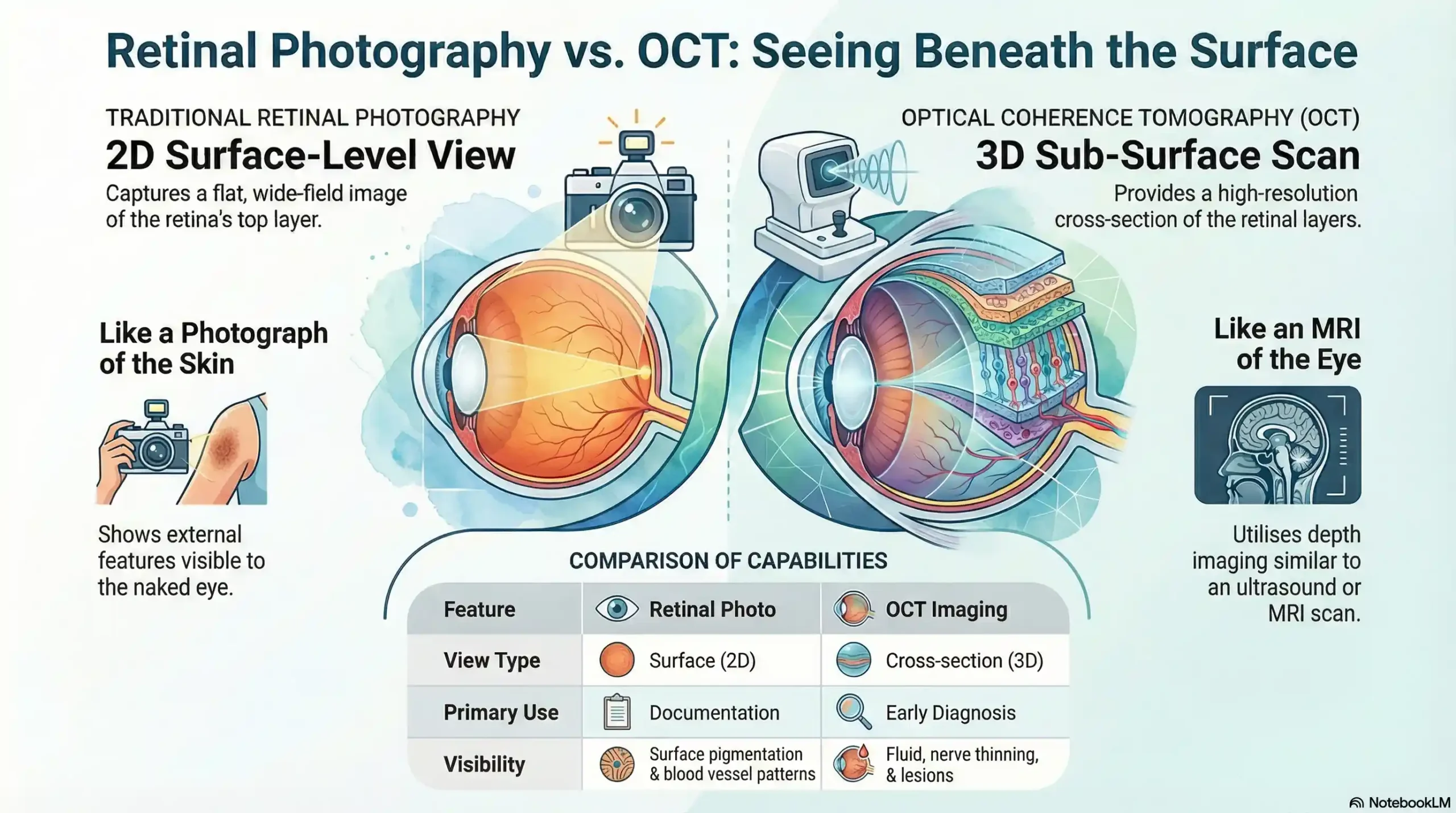
| Feature | Traditional Retinal Photo | Optical Coherence Tomography (OCT) |
| View Type | Surface-level (2D) | Sub-surface / Cross-section (3D) |
| Analogy | A photograph of the skin | An MRI or Ultrasound of the eye |
| What it Sees | Pigmentation & blood vessels | Fluid, nerve thinning, & hidden lesions |
| Benefit | Great for documentation | Essential for early diagnosis |
Protecting Your Child’s Future: OCT in Myopia Management
With children spending more time on digital devices, nearsightedness is on the rise. At Frame & Focus, we don’t just give your child stronger glasses every year; we use technology to actively manage the health of their growing eyes through Myopia Control.
How OCT helps your child:
-
Monitoring Eye Growth: As nearsightedness worsens, the eye often physically stretches. OCT allows us to monitor the thickness of the retina to ensure it isn’t becoming dangerously thin.
-
Personalised Treatment: Whether we recommend speciality contact lenses or MiSight therapy, the OCT provides the data needed to see if the treatment is working.
-
Safety First: The scan is non-invasive, takes seconds, and uses no radiation—making it perfectly safe for pediatric eye exams.
Resources and Citations
Professional Medical Organisations
American Academy of Ophthalmology – OCT Information
The American Academy of Ophthalmology provides comprehensive patient education materials about OCT technology, including what to expect during the procedure and how OCT images help diagnose various eye conditions. This resource offers evidence-based information reviewed by leading ophthalmologists.
American Optometric Association – Retinal Imaging Guidelines
The AOA provides detailed information about retinal imaging procedures, including OCT, and explains how these advanced diagnostic tools benefit patient care. This resource helps patients understand the role of OCT in comprehensive eye examinations.
If you would like to find out more about Optical Coherence Tomography, don’t hesitate to speak to our professional eye care team.
Secure Your Vision’s Future Today. Don’t wait for symptoms to appear. Join the Richmond neighbours who trust Dr Zaver’s “Straight Talk” approach and state-of-the-art OCT technology. Establish your eye health baseline today so we can protect your sight for a lifetime.
Schedule Your Comprehensive Exam & OCT Scan Or call us at (832) 930-7797
FAQs
-
An Optical Coherence Tomography (OCT) scan is a non-invasive test that uses light waves to create high-resolution, cross-sectional, 3D images of your retina and optic nerve.


