
The Importance of Retinal Imaging: What We Can Detect Early
Retinal imaging at Frame & Focus Eye Care in Richmond, Texas, provides a high-resolution map of your eye to detect silent conditions like glaucoma, diabetic retinopathy, and even early signs of Alzheimer’s disease. This non-invasive technology captures details up to five times more precisely than traditional exams, allowing for early intervention before permanent vision loss occurs.
Schedule Your Advanced Retinal Exam in Richmond
What is Retinal Imaging?
Retinal imaging is a sophisticated diagnostic technique that creates high-resolution digital photographs of the interior structures of your eye, specifically focusing on the retina, optic nerve, and blood vessels. Think of it as a detailed roadmap of your eye’s inner landscape—one that can reveal crucial information about both your vision and your general health.
This non-invasive procedure captures images that are up to 5 times more detailed than what can be observed during a traditional undilated eye examination. The technology creates a permanent digital record that becomes an invaluable part of your medical history, allowing our Richmond optometry team to track subtle changes over time that might otherwise go unnoticed.
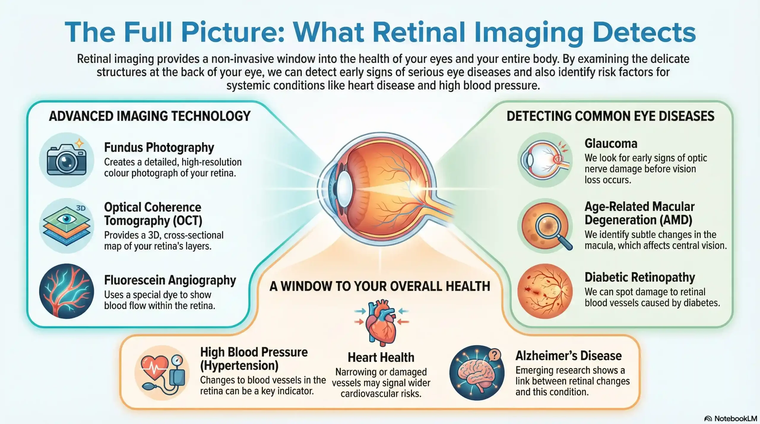
Advanced Retinal Imaging Technologies
Fundus Photography captures detailed color images of your retina’s surface, providing excellent visibility of blood vessels and helping us detect early signs of diabetic retinopathy and other vascular conditions.
Optical Coherence Tomography (OCT) uses light waves to create cross-sectional images of your retina’s layers, offering unprecedented detail for detecting macular degeneration, glaucoma, and other structural abnormalities.
Fluorescein Angiography uses contrast dye to highlight blood flow patterns in your retina, helping us identify circulation problems and vascular abnormalities in complex cases.
The Revolutionary Impact of Early Detection
The power of retinal imaging lies not just in what it can see, but in when it can see it. Many eye diseases progress gradually and silently, causing irreversible damage before symptoms become apparent. By the time a patient notices vision changes, the disease may have already advanced to stages where treatment options become limited and less effective.
Recent market research indicates that the global retinal imaging devices market was valued at approximately $3.74 billion in 2023, with continued growth driven by technological advances, including AI integration and improved accessibility. This expansion reflects the increasing recognition among healthcare professionals and patients of retinal imaging’s critical role in preventive eye care.
When we catch eye diseases in their earliest stages, treatment outcomes improve dramatically. Early intervention can often slow or stop disease progression, preserving your vision for years to come. This proactive approach aligns perfectly with our patient-centered philosophy at Frame & Focus Eye Care—we don’t just treat problems; we prevent them.
Eye Conditions We Can Detect Through Retinal Imaging
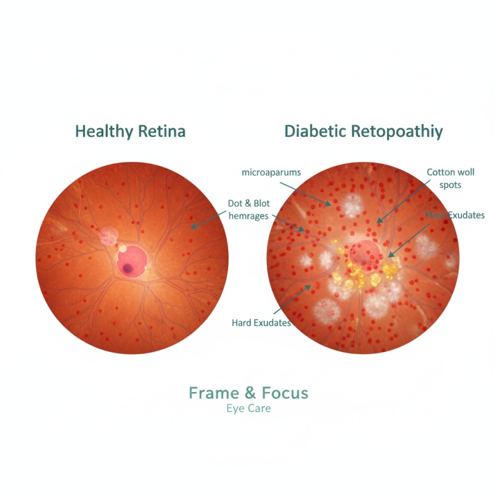
Diabetic Retinopathy: The Silent Vision Thief
According to the latest CDC data from 2021, diabetic retinopathy affects approximately 9.6 million Americans, with 1.84 million experiencing vision-threatening forms of the disease. This represents a prevalence rate of 26.43% among people with diabetes, with projections showing this could affect up to 14.6 million Americans by 2050.
Through retinal imaging, we can identify the earliest signs of diabetic retinopathy, including microaneurysms, dot and blot hemorrhages, hard exudates, and cotton wool spots. Our imaging technology allows us to detect these changes often years before symptoms develop, providing crucial time for you and your physician to optimize diabetes management and prevent vision loss.
Age-Related Macular Degeneration (AMD)
According to the latest CDC data, approximately 19.8 million Americans aged 40 and older were living with some form of age-related macular degeneration in 2019, with 1.49 million having vision-threatening late-stage AMD. AMD affects the macula, the central part of your retina responsible for sharp, detailed vision.
We can identify early AMD indicators such as drusen deposits, pigment changes in the retinal pigment epithelium, geographic atrophy in advanced dry AMD, and neovascularization in wet AMD. Detecting AMD early allows us to monitor progression closely and implement treatments that can significantly slow vision loss.
Glaucoma: The “Sneak Thief” of Sight
Glaucoma often develops without symptoms until significant vision loss has occurred. Through retinal imaging, particularly OCT technology, we can detect subtle changes in your optic nerve and retinal nerve fiber layer that may indicate glaucoma years before traditional testing methods.
Key glaucomatous changes we monitor include optic nerve head cupping, retinal nerve fiber layer thinning, ganglion cell complex reduction, and asymmetry between eyes.
Additional Conditions Detected
Retinal imaging also helps us identify signs of retinal tears, detachments, and blood vessel blockages that require immediate attention. High blood pressure often shows its first signs in the delicate blood vessels of your retina through arterial narrowing, cotton wool spots, flame-shaped hemorrhages, and hard exudates.
Beyond Eye Health: Systemic Health Detection
One of the most remarkable aspects of retinal imaging is its ability to provide insights into your systemic health. The retina contains the only blood vessels in your body that can be directly observed without invasive procedures, making it a unique window into your cardiovascular and neurological health.
Cardiovascular Disease Detection
Research has consistently shown strong correlations between retinal vascular changes and cardiovascular disease risk. Changes in retinal blood vessel caliber, tortuosity, and branching patterns can indicate increased stroke risk, coronary artery disease, hypertension, and atherosclerosis.
Neurological Condition Screening
Emerging research reveals exciting possibilities for using retinal imaging in the early detection of neurological conditions. A groundbreaking study published in NPJ Digital Medicine in October 2024 introduced the “Eye-AD” deep learning framework, which achieved remarkable accuracy in detecting early-onset Alzheimer’s disease using retinal imaging, with an Area Under the Curve (AUC) of 0.9355.
A comprehensive systematic review published in 2025 analyzed AI-assisted retinal imaging for neurodegenerative disease detection, showing an overall pooled AUC of 0.73 for detecting both Alzheimer’s and Parkinson’s diseases. This research represents a major breakthrough in using retinal imaging for neurological disease detection.
The Technology Behind Modern Retinal Imaging
The integration of artificial intelligence into retinal imaging represents one of the most significant advances in eye care. Three AI algorithms for diabetic retinopathy screening have received FDA approval: LumineticsCore (87.4% sensitivity, 89.5% specificity), EyeArt (96% sensitivity, 88% specificity), and AEYE-DS (92.6% sensitivity, 95.3% specificity).
Modern retinal imaging devices offer ultra-widefield imaging capturing up to 200 degrees of the retina, high-dynamic-range imaging for enhanced detail, and spectral domain OCT for precise layer analysis.
The Frame & Focus Eye Care Difference
At Frame & Focus Eye Care, we provide comprehensive retinal health assessments that become an integral part of your long-term health management strategy. Our retinal imaging process includes a detailed health history review, a comprehensive pre-imaging assessment by Dr. Sarah Zaver, high-resolution image capture, immediate analysis, and comprehensive reports explained in clear, understandable language.
The investment in retinal imaging provides early disease detection, treatment optimization, monitoring, peace of mind with a comprehensive assessment, and long-term medical records that benefit your family’s future eye care.
Understanding the Investment
Retinal imaging typically costs between $25-60 per session, with most practices charging in the $35-40 range. According to current 2025 data, pricing varies based on the technology used, your geographic location, and your insurance coverage. While most vision insurance plans don’t cover this elective procedure, many medical insurance plans may cover retinal imaging when medically indicated.
When considering the cost, it’s important to weigh the expense of preventive screening against potential future treatment costs. Early detection of eye diseases can prevent more expensive interventions later and preserve irreplaceable vision.
Who Should Consider Retinal Imaging?
High-risk individuals particularly benefit from retinal imaging, including diabetics and pre-diabetics, individuals with high blood pressure, those with a family history of eye disease, people age 40 and above, and those with previous eye injuries or surgeries.
Even if you’re not in a high-risk category, retinal imaging offers significant value for preventive care. The detailed baseline images we create today become invaluable references for detecting future changes, no matter how subtle.
What to Expect During Your Appointment
Retinal imaging is completely painless and non-invasive. You’ll sit comfortably in front of our imaging device, see a bright flash of light as the camera captures the image, and receive immediate review of images with Dr. Zaver explaining what she sees and what it means for your health.
The entire process typically takes 5-10 minutes, with no recovery time needed. Unlike traditional dilated exams, retinal imaging doesn’t affect your vision afterward, allowing you to drive immediately and return to normal activities without restriction.
References and Additional Resources
For readers interested in learning more about retinal imaging research and statistics, we recommend these authoritative sources that informed this article:
- CDC Vision and Eye Health Surveillance System (VEHSS) – Diabetic Retinopathy Prevalence Data.
The Centers for Disease Control and Prevention’s comprehensive database provides the latest 2021 prevalence estimates for diabetic retinopathy, showing 9.6 million Americans affected. - NPJ Digital Medicine – Early Detection of Dementia Through Retinal Imaging and AI. This groundbreaking October 2024 study demonstrates how retinal imaging can detect early-onset Alzheimer’s disease with 93.55% accuracy using AI technology.
- JAMA Ophthalmology – Prevalence of Diabetic Retinopathy in the US in 2021. This comprehensive study provides the most current prevalence estimates of diabetic retinopathy by demographic factors across US counties and states.
Your Next Steps: Schedule Your Comprehensive Eye Examination
At Frame & Focus Eye Care, we believe that proactive eye care is the foundation of lifelong vision health. Retinal imaging represents our commitment to providing you with the most advanced, comprehensive care available. Through our state-of-the-art retinal imaging technology, we can detect potential problems before they threaten your vision.
Why Choose Frame & Focus Eye Care? Dr. Sarah Zaver brings over a decade of experience serving the Houston area. We utilize the latest imaging equipment for detailed assessments, we provide patient-centered care with clear explanations and personalized care plans, and we offer a convenient Richmond location for residents of Richmond, Pecan Grove, Sugar Land, Rosenberg, and Fulshear.
Contact Frame & Focus Eye Care:
- Address: 18310 W Airport Blvd #900, Richmond, TX 77407
- Online Scheduling: Available for your convenience
- Same-Day Appointments: Available for urgent needs
Don’t wait for symptoms to develop. Take a proactive step toward protecting your vision and overall health by scheduling a comprehensive eye examination with retinal imaging today.
At Frame & Focus Eye Care, we’re more than just an eye clinic—we’re your partners in maintaining lifelong vision health. Dr. Sarah Zaver and our experienced team are committed to providing exceptional, personalized care that combines the latest technology with the human touch that makes you feel like family.
FAQs
-
Digital retinal imaging captures a high-resolution photo of the back of your eye. It is vital for detecting diseases like glaucoma and diabetic retinopathy before they cause permanent vision loss.
Please note: None of the above should be considered medical advice. If you have any concerns about your vision, please contact us immediately or consult your primary care provider.


