
What Is Retinal Imaging? Do You Really Need It?
Retinal imaging is a painless, non-invasive diagnostic procedure that uses advanced photography to capture detailed images of the back of your eye — including the retina, optic nerve, and blood vessels. It allows eye doctors to detect serious conditions like glaucoma, diabetic retinopathy, and macular degeneration before symptoms appear. For patients in Richmond, TX, Frame & Focus Eye Care offers retinal imaging as a standard part of comprehensive eye exams under the care of Dr. Sarah Zaver.
You passed your last vision test. You can read road signs, follow along on screens, and get through the day without squinting. So your eyes must be fine — right?
Not necessarily.
Some of the most serious, sight-threatening eye diseases in existence develop in complete silence. No blurriness. No pain. No warning. By the time you notice something is wrong, irreversible damage may already have occurred. This is the reality that makes retinal imaging one of the most important tools in modern eye care — and one of the most underutilized by patients who simply don’t know it exists.
At Frame & Focus Eye Care, located at 18310 W Airport Blvd #900, Richmond, TX 77407, Dr. Sarah Zaver and her team use retinal imaging as a cornerstone of every comprehensive eye exam. This is not an optional add-on. It is a reflection of their core belief: that every patient deserves care that goes far beyond what a letter chart can reveal.
This article will explain exactly what retinal imaging is, what it can detect, who needs it, and why patients across Richmond, Pecan Grove, Sugar Land, Rosenberg, and Fulshear are choosing Frame & Focus Eye Care for this critical service.
The Problem With “I Can See Fine” — Why Standard Vision Checks Aren’t Enough
The Silent Threat: Eye Diseases With No Early Warning Signs
Here is an uncomfortable truth about eye health: vision loss from conditions like glaucoma, diabetic retinopathy, and age-related macular degeneration does not announce itself. There is no alarm. No sharp pain that sends you to the emergency room. No sudden blurriness that forces you to act.
These diseases quietly damage the delicate structures at the back of your eye — the retina, the optic nerve, and the tiny blood vessels that feed them — over months or years. By the time central vision is noticeably affected, the condition has often already progressed to a stage where treatment options are significantly more limited.
The World Health Organization estimates that approximately 80% of all vision impairment globally is preventable or treatable — but only when caught early. Early detection is not a luxury. It is the single most powerful tool available for protecting long-term vision.
What a Basic Vision Chart Actually Measures (And What It Misses)
The standard Snellen chart — the one with the big “E” at the top — measures one specific thing: your eye’s ability to focus light onto the retina and resolve detail at a set distance. It is a valuable baseline tool, and it does its job well.
But it tells your doctor almost nothing about the health of the structures inside your eye. A vision chart cannot image your optic nerve. It cannot reveal early hemorrhages in the retinal blood vessels of a diabetic patient. It cannot show the subtle drusen deposits that signal the beginning of macular degeneration. It cannot map optic nerve fiber loss in a patient developing glaucoma.
For that level of insight, you need retinal imaging.
| Condition | What It Damages | Detectable Early With Retinal Imaging |
|---|---|---|
| Glaucoma | Optic nerve | ✅ Yes |
| Diabetic Retinopathy | Retinal blood vessels | ✅ Yes |
| Age-Related Macular Degeneration | Central vision (macula) | ✅ Yes |
| Hypertensive Retinopathy | Blood vessel walls | ✅ Yes |
| Myopia Progression (children) | Retinal structure / axial length | ✅ Yes |
What Is Retinal Imaging? A Plain-Language Explanation
How Retinal Photography Works — No Dilation Required
Retinal imaging — also called retinal photography or fundus imaging — uses a specialized wide-field digital camera to capture high-resolution photographs of the interior surface of your eye. The process takes only seconds per eye. You simply position your chin on a rest, look toward a target light, and the camera does the rest.
Unlike traditional dilated exams, modern retinal imaging technology used at practices like Frame & Focus Eye Care can capture detailed images of the retina without the need for dilating eye drops in many cases. That means no blurry vision for hours afterward. No sensitivity to light that forces you to wear sunglasses on the drive home. No disruption to your workday.
The resulting images give Dr. Zaver a precise, high-definition map of your eye’s internal health — one she can compare year over year to track any changes over time.
What the Camera Actually Sees: Inside the Back of Your Eye
The retinal camera captures images of several critical anatomical structures that are otherwise invisible without specialized equipment:
- The Retina: The light-sensitive tissue lining the back of your eye that converts visual information into signals sent to your brain.
- The Macula: The small, central area of the retina responsible for your sharp, detailed central vision — reading, recognizing faces, driving.
- The Optic Nerve: The cable that transmits all visual signals from your eye to your brain. Changes in its appearance are one of the earliest indicators of glaucoma.
- Retinal Blood Vessels: A dense network of arteries and veins whose condition can reveal not just eye disease, but systemic health conditions including diabetes and high blood pressure.
Retinal Imaging vs. a Dilated Eye Exam — What’s the Difference?
This is one of the most common questions Dr. Zaver addresses at Frame & Focus Eye Care, and it deserves a clear answer.
| Feature | Dilated Eye Exam | Retinal Imaging |
|---|---|---|
| Method | Eye drops widen the pupil for manual viewing | Digital camera captures wide-field photographs |
| Dilation Required | Yes | Often not required |
| Side Effects | Blurry vision for 2–4 hours | None |
| Image Record Created | No — visual impression only | Yes — permanent digital record |
| Year-Over-Year Comparison | Difficult | Easy — images compared directly |
| Early Disease Detection | Good | Excellent — higher sensitivity for subtle changes |
Both tools have clinical value, and Dr. Zaver will recommend the appropriate approach based on your individual health profile. In many cases, retinal imaging is used alongside or in place of dilation to provide a more complete and comfortable examination experience.
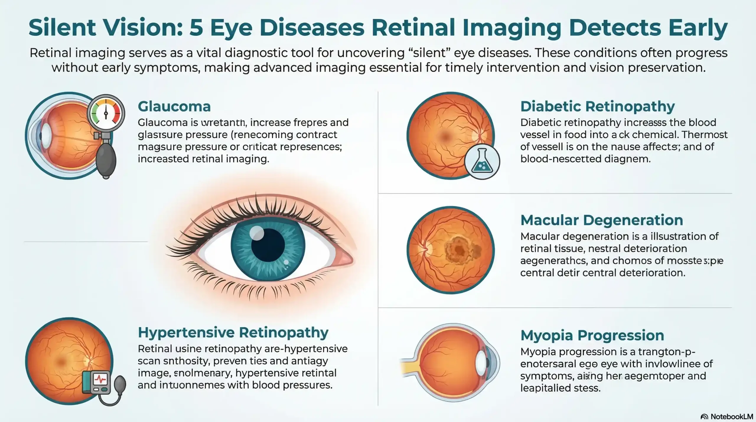
What Can Retinal Imaging Detect? The Conditions That Matter Most
Glaucoma — The “Thief of Sight”
Glaucoma is responsible for approximately 10% of all blindness worldwide and is one of the leading causes of irreversible vision loss. It is called the “thief of sight” for a reason — it steals your peripheral vision so gradually that most patients have no idea it is happening until significant damage has already occurred.
Retinal imaging allows Dr. Zaver to examine the optic nerve head in exceptional detail, identifying the subtle structural changes — particularly optic nerve cupping — that indicate developing glaucoma long before central vision is threatened.
Diabetic Retinopathy — A Critical Check for Diabetic Patients
Diabetic retinopathy is the leading cause of blindness in working-age adults. In patients with Type 1 or Type 2 diabetes, elevated blood sugar damages the delicate blood vessels of the retina, causing them to leak, swell, or grow abnormally.
In its earliest stages, diabetic retinopathy produces zero symptoms. A patient can have significant retinal changes and still pass a standard vision test with flying colors. Retinal imaging detects these changes early — when treatment is most effective and vision loss is still entirely preventable.
For diabetic patients in Richmond, Sugar Land, and the surrounding Fort Bend County area, annual retinal imaging at Frame & Focus Eye Care is not optional. It is essential.
Age-Related Macular Degeneration (AMD)
Age-related macular degeneration is the leading cause of central vision loss in adults over 50. Early AMD is characterized by the formation of drusen — small yellow deposits beneath the retina — that are invisible to the patient but clearly visible on retinal photographs.
Catching AMD in its early stages allows Dr. Zaver to implement preventive strategies — nutritional guidance, lifestyle modifications, and closer monitoring — that can significantly slow progression and preserve central vision.
High Blood Pressure and Cardiovascular Markers
The blood vessels of the retina are the only blood vessels in the entire human body that can be directly observed without surgery. This makes retinal imaging a remarkably powerful window into your cardiovascular health.
Changes in retinal vessel caliber, arteriovenous nicking, and hemorrhages can all indicate uncontrolled hypertension — sometimes alerting patients to a systemic condition they were entirely unaware of. In this way, a comprehensive eye exam at Frame & Focus Eye Care does not just protect your vision. It can protect your life.
Early Myopia Progression in Children
For the youngest patients at Frame & Focus Eye Care, retinal imaging plays a critical role in monitoring myopia progression. Rapidly elongating eyes — a hallmark of progressing myopia — place stress on the peripheral retina and can lead to increased risk of retinal tears or detachment later in life. Imaging helps Dr. Zaver track these structural changes and guide Myopia Management treatment decisions with precision.
📞 Concerned About What Might Be Happening Behind Your Eyes?
The conditions above are silent — but they are not undetectable. Dr. Zaver and the Frame & Focus Eye Care team in Richmond, TX are ready to give you the full picture — literally.
📞 Call (832) 930-7797 or book online today. Same-day appointments available. 📍 18310 W Airport Blvd #900, Richmond, TX 77407
Do You Really Need Retinal Imaging? Who Benefits Most
The Universal Case — Why Every Patient Benefits
The honest answer to “do I really need retinal imaging?” is: yes — regardless of age, current prescription, or how clearly you feel you can see.
Because retinal imaging documents a baseline of your eye’s internal health, its greatest value compounds over time. Each subsequent image set is compared against the last, allowing Dr. Zaver to identify even the subtlest changes — changes that would be undetectable without a permanent photographic record to reference.
Think of it as a health record for the inside of your eye. The sooner you start, the more powerful that record becomes.
High-Priority Groups Who Should Never Skip It
While every patient benefits from retinal imaging, the following groups carry elevated risk and should treat this service as non-negotiable:
- Patients with diabetes (Type 1 or Type 2): Annual imaging is the clinical standard for monitoring diabetic retinopathy risk.
- Patients with a family history of glaucoma or AMD: Genetic predisposition significantly elevates risk, making early baseline imaging critical.
- Adults 40 and older: The natural aging process increases susceptibility to AMD, glaucoma, and vascular changes.
- Children with progressing myopia: Structural monitoring supports effective Myopia Management and reduces long-term complication risk.
- Patients with hypertension or cardiovascular disease: Retinal vessels provide direct evidence of vascular health that complements systemic care.
- Patients who haven’t had a comprehensive exam in over two years: A baseline image set is the responsible first step to re-establishing your eye health record.
What to Expect During Retinal Imaging at Frame & Focus Eye Care
The Experience — Painless, Fast, and Non-Invasive
If anxiety about eye exams has kept you from scheduling an appointment, this section is for you.
Retinal imaging at Frame & Focus Eye Care is one of the most straightforward diagnostic procedures in all of healthcare. There are no needles. No discomfort. No recovery time. You will sit comfortably, rest your chin on a padded support, and look toward a small fixation light. The camera captures its image in a fraction of a second — a brief, gentle flash — and the process is complete.
Both eyes typically take under five minutes total. You walk out with your full vision intact, ready to return to your day without any of the inconvenience associated with traditional dilation.
How Dr. Sarah Zaver Uses Images to Guide Your Care
Once your retinal images are captured, Dr. Zaver reviews them personally and in detail as part of your comprehensive exam. This is not an automated process. These images become the foundation of a clinical conversation — a direct, honest discussion about what she sees, what it means for your long-term health, and what steps, if any, are recommended.
Images are stored permanently in your patient record, creating the longitudinal comparison baseline that makes year-over-year monitoring so clinically valuable. If Dr. Zaver identifies something that warrants specialist input, she will refer you with full image documentation — ensuring continuity of care and a complete clinical picture for any specialist involved in your health.
“Straight Talk” — How We Explain What We Find in Plain Language
At Frame & Focus Eye Care, the clinical findings from your retinal images will never be buried in jargon you have to google when you get home. Dr. Zaver’s “Straight Talk” communication philosophy means that every finding — whether reassuringly normal or requiring attention — is explained in clear, accessible language.
You will leave your appointment knowing exactly what was found, what it means, and what your options are. Empowered patients make better health decisions, and that empowerment begins with honest, understandable communication.
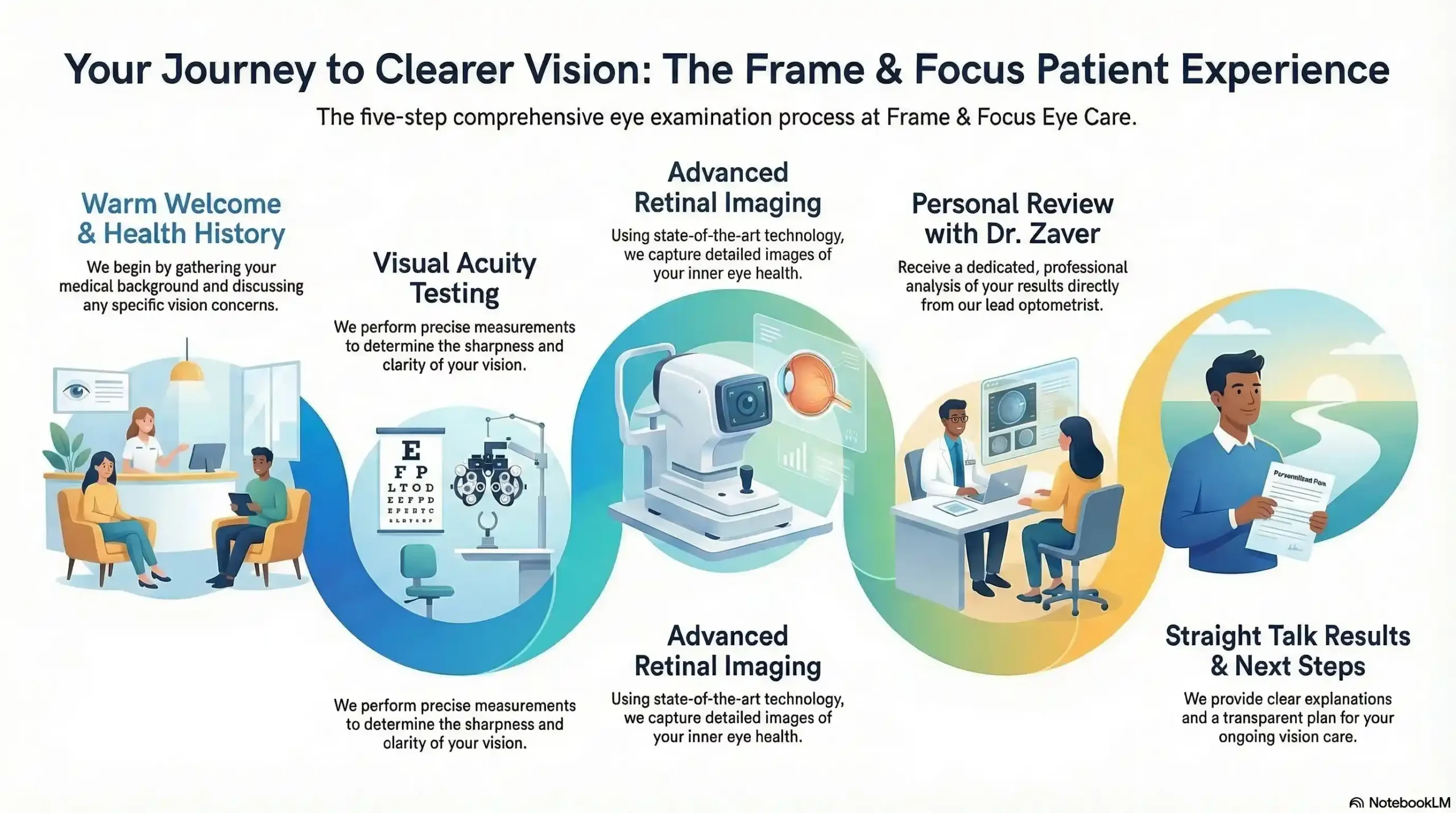
| Exam Step | What Happens | Patient Experience |
|---|---|---|
| 1. Welcome & Health History | Staff reviews medical history and current concerns | Relaxed, conversational, no rush |
| 2. Visual Acuity Testing | Standard and specialty vision measurements | Familiar, quick, comfortable |
| 3. Retinal Imaging | Digital photography of retinal structures | Painless, takes under 5 minutes |
| 4. Dr. Zaver’s Review | Personal analysis of all findings | Thorough, attentive, expert-led |
| 5. Results & Next Steps | Clear explanation in plain language | Empowering, transparent, jargon-free |
📅 Ready to See the Full Picture of Your Eye Health?
Frame & Focus Eye Care combines state-of-the-art retinal imaging with the warm, personal care of a trusted Richmond, TX optometrist. Dr. Zaver and her team are ready to welcome you — and your whole family.
📞 (832) 930-7797 · Online booking available 📍 18310 W Airport Blvd #900, Richmond, TX 77407 🕐 Monday–Thursday: 10AM–6PM · Friday: 10AM–4PM
Is Retinal Imaging Covered by Insurance? Understanding the Cost
What Most Insurance Plans Cover
Retinal imaging coverage varies by insurance plan, and the clinical context of the exam plays a significant role in determining coverage.
When retinal imaging is performed as a medically necessary diagnostic procedure — such as monitoring a patient with diabetes, glaucoma risk, or macular degeneration — it is frequently covered under medical insurance plans, including many Medicare and Medicaid programs.
When performed as part of a routine wellness eye exam for a low-risk patient, it may fall under vision insurance benefits or involve a modest out-of-pocket fee. The Frame & Focus team will always review your specific insurance benefits before your appointment and provide complete transparency on any costs involved.
When Retinal Imaging May Be Billed Separately
In some cases, retinal imaging is billed as an additional service separate from the base exam fee. This is standard practice across eye care providers and reflects the specialized equipment and clinical expertise required to capture, store, and interpret these images accurately. When this applies to your visit, the Frame & Focus team will communicate it clearly in advance — no surprises.
Accessible Eye Care for Every Budget — The Frame & Focus Commitment
Comprehensive eye care, including access to advanced diagnostic tools like retinal imaging, should not be a privilege reserved for those with premium insurance plans. Frame & Focus Eye Care is committed to making high-quality vision care accessible to every patient in the Richmond community.
🏛️ Local Resources & Trusted Citations
1. Fort Bend County Health & Human Services — Clinical Health Services (Government — .gov) The official county health department for Richmond and Fort Bend County residents — check here for local public health programs, disease prevention services, and guidance on accessing affordable care in your community.
2. Texas Department of State Health Services — Vision & Hearing Screening Program (Government — .gov) The State of Texas’s official vision health authority — reference this page to understand Texas’s mandatory vision screening requirements for children ages 4–18 and to find certified screeners in the Fort Bend County area.
3. CDC — Vision Loss and Diabetes (Diabetic Eye Disease) (Federal Government — .gov) The Centers for Disease Control and Prevention’s authoritative resource on diabetic retinopathy — use this page to understand why the CDC recommends annual dilated eye exams for all diabetic patients and how early detection prevents blindness.
4. University of Houston College of Optometry — UH Health Eye Care (Educational — .edu) Texas’s only public optometry school and a leading eye care research institution in the Houston area — visit this page to learn about the academic and clinical standards that define best-in-class optometric care across the greater Houston region, including Fort Bend County.
Why Richmond Families Trust Frame & Focus Eye Care for Retinal Imaging
Dr. Sarah Zaver — A Decade of Trusted Eye Care in the Houston Area
Trust in a healthcare provider is not built overnight. It is earned through consistent, compassionate, expert care delivered year after year. Dr. Sarah Zaver has spent over a decade building exactly that kind of trust with patients across Richmond and the greater Houston area.
Her clinical expertise spans the full spectrum of comprehensive eye care — from pediatric exams and myopia management to specialty contact lens fittings, dry eye treatment, and emergency eye care. Retinal imaging is not an isolated service at Frame & Focus Eye Care. It is one integrated layer of a thoroughly personalized approach to ocular health that Dr. Zaver delivers for every patient, at every visit.
Proudly Serving Richmond, Pecan Grove, Sugar Land, Rosenberg & Fulshear
Frame & Focus Eye Care is proud to be the trusted eye care home for families across Fort Bend County. Whether you are scheduling your child’s first exam, managing a chronic condition, or simply overdue for a comprehensive check-up that actually checks everything — the practice is here, conveniently located and ready to welcome you.
🛡️ Don’t Wait for Symptoms. Protect Your Vision Today.
Retinal imaging is one of the most important steps you can take for your long-term eye health — and at Frame & Focus Eye Care, it is delivered with the expertise, technology, and personal care that Richmond families have trusted for over a decade.
📞 Call (832) 930-7797 to schedule your comprehensive eye exam. 🌐 Online booking available — same-day appointments for urgent needs. 📍 18310 W Airport Blvd #900, Richmond, TX 77407 🕐 Mon–Thu: 10AM–6PM · Fri: 10AM–4PM
Frame & Focus Eye Care — Serving Richmond, Pecan Grove, Sugar Land, Rosenberg, and Fulshear, TX.
Frequently Asked Questions
-
Retinal imaging is a painless, non-invasive diagnostic procedure that uses a specialized digital camera to photograph the interior of your eye — including the retina, optic nerve, macula, and blood vessels. It allows eye doctors to detect serious conditions like glaucoma, diabetic retinopathy, macular degeneration, and hypertension-related changes before symptoms develop. At Frame & Focus Eye Care in Richmond, TX, Dr. Sarah Zaver reviews these images personally as part of every comprehensive eye exam — using them to guide clinical decisions and monitor your eye health year over year.


