
Retinal Imaging vs. Eye Dilation: What’s the Real Difference?
Retinal imaging uses a high-resolution digital camera to photograph the back of your eye without drops, while eye dilation uses medicated drops to widen the pupil for a hands-on examination. Both detect serious conditions like glaucoma, macular degeneration, and diabetic retinopathy — but retinal imaging lets you drive home afterward, stores a digital record for future comparison, and causes no side effects. In many routine exams, retinal imaging is sufficient; your eye doctor will advise when dilation is still the better choice.
Why This Question Matters More Than You Think
You’ve just settled into the exam chair at your eye appointment. Before the doctor comes in, a staff member hands you a clipboard. Near the bottom, you notice a question you weren’t expecting: “Would you like retinal imaging added to your exam today for an additional fee, or would you prefer dilation?”
If you’ve ever stared at that question without knowing what to say, you’re not alone. Most patients have never been given a clear, jargon-free explanation of what either procedure actually involves — and without that context, the choice can feel overwhelming.
At Frame & Focus Eye Care in Richmond, TX, we believe you should never have to make a healthcare decision without fully understanding your options. That’s our “Straight Talk” promise. So let’s break this down clearly — no optometry jargon, just real answers.
Understanding the Back of Your Eye — What Doctors Are Looking For
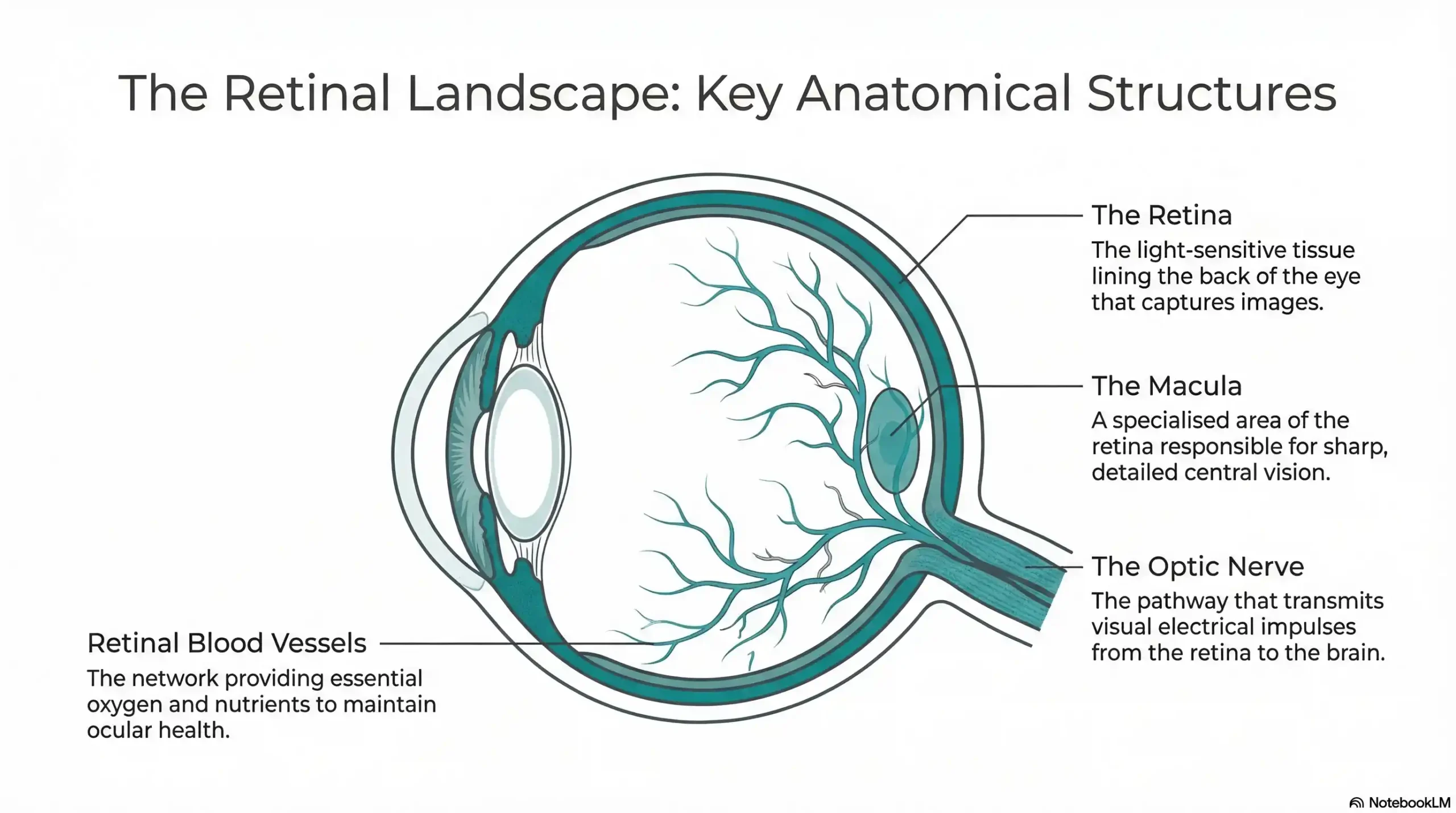
Both retinal imaging and dilation are designed to give your eye doctor a clear view of the structures at the back of your eye. To understand why that matters, it helps to know what those structures are and what they do.
The retina is the light-sensitive layer of tissue lining the inside of your eye. It converts what you see into electrical signals that travel to your brain. The optic nerve carries those signals from your eye to your brain — and its appearance can reveal early signs of glaucoma. The macula is the central part of the retina responsible for sharp, detailed vision. The blood vessels running through the retina can reflect the health of your cardiovascular system and flag conditions like diabetes and high blood pressure.
Here’s the critical point: many of the most serious eye conditions — glaucoma, macular degeneration, diabetic retinopathy — produce no noticeable symptoms in their early stages. By the time you notice something is wrong, significant, irreversible damage may already have occurred. That’s precisely why examining the back of the eye during a routine exam is so important.
| Eye Structure | Why It Matters |
|---|---|
| Retina | Converts light to signals; affected by diabetic retinopathy and retinal detachment |
| Optic Nerve | Transmits vision to brain; damage is a key glaucoma indicator |
| Macula | Responsible for central vision; degeneration leads to progressive vision loss |
| Blood Vessels | Reflects cardiovascular health; can reveal hypertension and diabetes |
What Is Eye Dilation? The Classic Method Explained
How Dilation Works
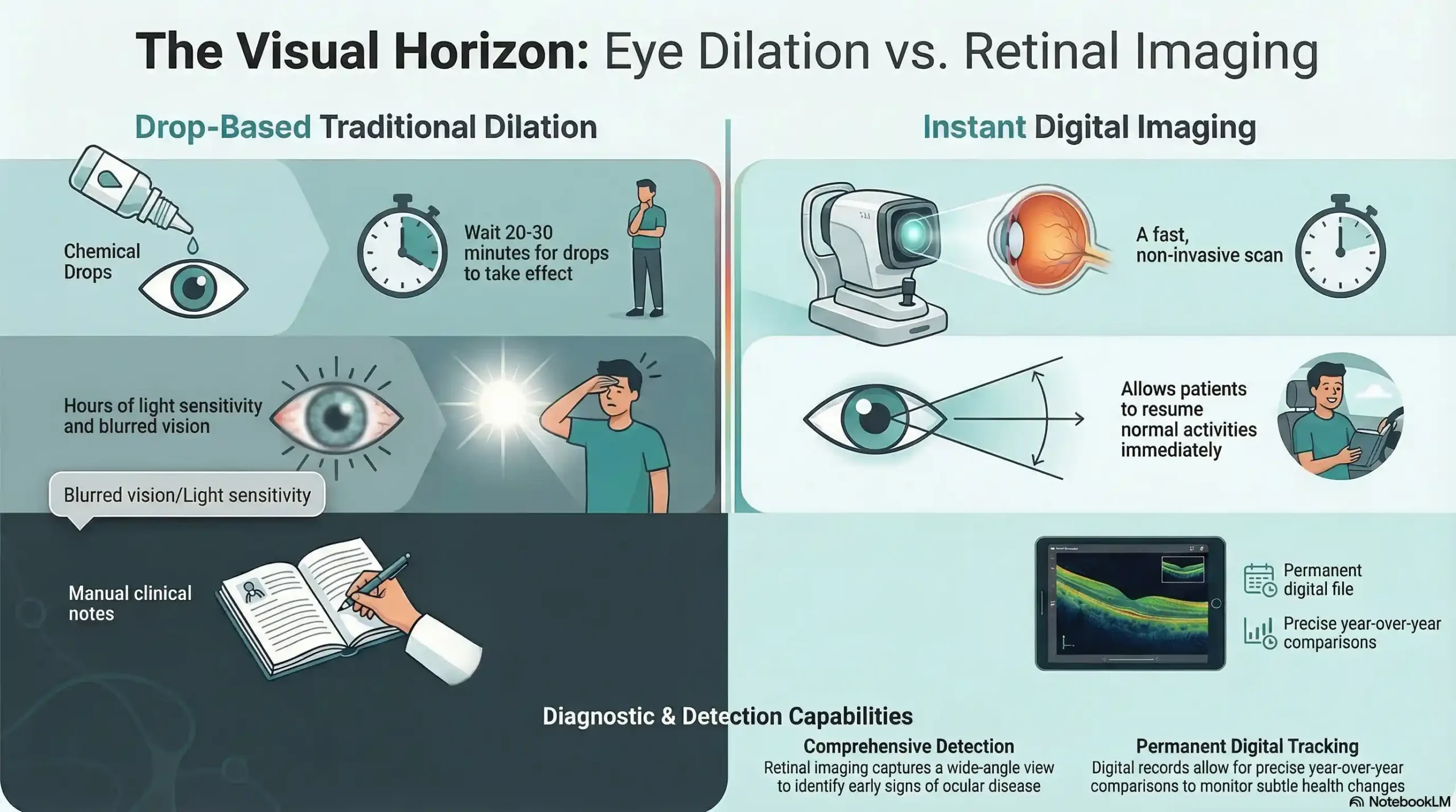
Eye dilation has been the standard method for examining the back of the eye for decades. Your eye doctor places medicated drops — known as mydriatic drops — into your eyes. These drops cause the pupil to expand significantly, typically to around 6 to 8 millimeters in diameter, compared to its natural resting size of 2 to 4 millimeters. This enlarged opening gives the doctor a direct, wide-angle view of the retina, optic nerve, and surrounding structures using a handheld light and lens.
What Dilation Can Detect
Dilation is a powerful diagnostic tool. When performed correctly, it allows your doctor to examine the full peripheral retina and identify a wide range of conditions, including:
- Glaucoma — via optic nerve assessment
- Diabetic retinopathy — by examining retinal blood vessels for leakage or abnormal growth
- Macular degeneration — early drusen deposits visible before vision loss begins
- Retinal tears or detachment — often asymptomatic until advanced
- Hypertensive retinopathy — changes in blood vessels linked to high blood pressure
The Trade-Offs Patients Experience
Dilation is highly effective, but it comes with side effects that many patients find inconvenient — and sometimes anxiety-inducing. Here’s what to expect:
- Blurry near vision lasting 2 to 4 hours (sometimes up to 6 hours for lighter-colored eyes)
- Significant light sensitivity — sunglasses are strongly recommended
- Difficulty driving — many patients need to arrange a ride home
- A slightly unusual appearance due to enlarged pupils
These effects are temporary and harmless, but they are a real consideration for patients with busy schedules, active children, or demanding work environments.
What Is Retinal Imaging? The Modern Alternative
How Retinal Photography Works
Retinal imaging — sometimes called retinal photography or optomap imaging — uses a high-resolution digital camera to capture a wide-field photograph of the back of your eye. The process is quick, non-invasive, and in most cases requires no drops at all. You simply look into the imaging device for a brief moment while the camera captures a detailed image of your retina, optic nerve, and blood vessels.
The result is an immediate, high-quality digital image that is stored in your patient file, creating a permanent visual record of your eye health over time.
What Retinal Imaging Can Detect
Modern retinal imaging is clinically validated to detect the same range of conditions as dilation, including glaucoma, macular degeneration, diabetic retinopathy, and retinal detachment. But it offers one significant additional advantage: the ability to compare images year over year.
Because your retinal photograph is stored digitally, your eye doctor can place this year’s image alongside last year’s and identify even subtle changes that might not yet be causing symptoms. This makes retinal imaging an exceptionally powerful tool for early detection and long-term monitoring of chronic eye conditions.
The Patient Experience Advantage
Here is where retinal imaging becomes a clear winner for many patients: there are no side effects. No blurry vision. No light sensitivity. No need to arrange a driver. You can walk into Frame & Focus Eye Care on your lunch break, have a comprehensive exam with retinal imaging, and drive yourself back to the office without missing a beat.
This comfort-forward experience is a reflection of our commitment to care that fits your life — not the other way around.
| Feature | Eye Dilation | Retinal Imaging |
|---|---|---|
| Requires drops | Yes — mydriatic eye drops | No (in most cases) |
| Blurry vision after exam | Yes — 2 to 4 hours | No |
| Can drive immediately after | Not recommended | Yes |
| Detects major eye diseases | Yes | Yes |
| Digital record kept | No | Yes — stored for comparison |
| Year-over-year change tracking | Limited | Yes — highly effective |
| Typical out-of-pocket cost | Usually insurance covered | Small additional fee typical |
| Patient comfort | Moderate discomfort / sensitivity | High — no side effects |
💬 Not sure which option is right for you? The team at Frame & Focus Eye Care is happy to walk you through both — in plain language, no jargon. 📞 Call us: (832) 930-7797 📅 Or book online — same-day appointments available.
Can Retinal Imaging Replace Dilation Entirely?
This is one of the most common questions patients ask — and the honest answer is: sometimes yes, sometimes no.
For the majority of routine annual exams in healthy adults with no known risk factors, wide-field retinal imaging provides sufficient information for a thorough assessment. In these cases, many optometrists — including our team at Frame & Focus — may recommend imaging as the primary method, reserving dilation for cases where a closer or more three-dimensional view is needed.
Dilation remains the preferred or required option in scenarios such as:
- Patients with diabetes who require a complete peripheral retinal evaluation
- Cases where a retinal tear or detachment is suspected
- Patients with a strong family history of glaucoma requiring a detailed optic nerve view
- Situations where imaging reveals something that warrants immediate follow-up
- Pediatric patients in certain clinical situations
The key takeaway: this is not a decision you need to make alone. Dr. Sarah Zaver will evaluate your individual health history, discuss your options clearly, and recommend the approach that genuinely serves your long-term eye health — not just a default protocol.
Does Insurance Cover Retinal Imaging?
Cost is understandably one of the biggest questions patients have when retinal imaging is offered. Here’s a straightforward breakdown:
Standard vision insurance: Retinal imaging is typically considered an elective add-on and is not automatically covered by standard vision plans. The usual out-of-pocket cost ranges from approximately $25 to $45, depending on the provider and technology used.
Medical insurance: If retinal imaging is deemed medically necessary — for example, if you have diabetes, a known history of retinal disease, or other qualifying conditions — your medical insurance may provide coverage. This is worth confirming before your appointment.
Eye dilation: Dilation is almost always covered as part of a standard comprehensive eye exam under vision insurance plans.
At Frame & Focus Eye Care, we believe in transparent, no-surprise pricing. Our team is happy to review your specific insurance benefits before your exam so you understand exactly what to expect — and how to get the most value from your coverage.
💬 We help you maximize your insurance benefits — no surprises. Schedule your comprehensive eye exam at our Richmond, TX office. 📍 18310 W Airport Blvd #900, Richmond, TX 77407 📞 (832) 930-7797 | Same-day appointments available.
What Frame & Focus Eye Care Recommends — And Why
With over a decade of experience serving the Richmond, TX community, Dr. Sarah Zaver has guided thousands of patients through exactly this decision. Her approach is straightforward: no upselling, no one-size-fits-all protocols — just personalized guidance rooted in your individual needs.
At Frame & Focus, we have integrated retinal photography into our comprehensive eye exams because we believe in the power of technology to catch what the naked eye might miss — and to create a longitudinal record of your eye health that becomes more valuable with every passing year.
As Dr. Zaver explains to her patients: “Think of retinal imaging as taking a photograph of the back of your eye. Every year we compare it to the year before. We’re not just looking for problems today — we’re watching for change over time. That’s where early detection really happens.”
This commitment to using state-of-the-art technology in a warm, approachable environment has earned Frame & Focus Eye Care a 4.9-star Google rating from over 315 patient reviews. Patients consistently praise the practice for its thoroughness, clarity of communication, and genuinely welcoming atmosphere.
We proudly serve patients from Richmond, Pecan Grove, Sugar Land, Rosenberg, and Fulshear — and we’d be honored to be your family’s trusted eye care partner.
🔗 Local Resources & Citations
Here are 4 authoritative, non-competitor entities relevant to Retinal Imaging, Eye Exams & Diagnostic Testing for Richmond / Fort Bend County, TX:
1. National Eye Institute (NEI) — National Institutes of Health A U.S. federal (.gov) health agency providing clinically validated information on conditions detected by retinal imaging, including glaucoma, macular degeneration, and diabetic retinopathy — ideal for patients who want to understand what their exam results mean.
2. Fort Bend County Public Health Department The official (.gov) county health authority for Richmond, TX — references here reinforce local relevance and signal to Google that Frame & Focus serves a recognized Fort Bend County community health need.
3. Texas Optometry Board — State of Texas The official (.gov) state licensing board that regulates optometrists in Texas, including Richmond — citing this establishes that Frame & Focus and Dr. Zaver operate under verified state professional standards.
4. University of Houston College of Optometry A leading Houston-area (.edu) optometry institution whose published patient education resources on retinal health and comprehensive eye exams lend strong academic authority to supporting content on this topic.
Protect Your Vision — Schedule Your Eye Exam in Richmond, TX
The most important thing to take away from this discussion is simple: whether you choose retinal imaging, dilation, or a combination of both — the exam itself is what matters most.
Many of the conditions that threaten your long-term vision are completely silent in their early stages. Glaucoma. Macular degeneration. Diabetic retinopathy. They don’t announce themselves with pain or obvious symptoms. They’re found by a thorough eye doctor using the right tools at the right time.
Don’t wait for something to feel wrong. Schedule your comprehensive eye exam with Frame & Focus Eye Care today and leave with confidence — knowing your vision and ocular health have been fully prioritized and customized to your unique needs.
📅 Ready to See Clearly? Book Your Exam Today.
Frame & Focus Eye Care | Richmond, TX 📍 18310 W Airport Blvd #900, Richmond, TX 77407 📞 (832) 930-7797 🕐 Mon–Thu: 10AM–6PM | Fri: 10AM–4PM
Proudly serving Richmond, Pecan Grove, Sugar Land, Rosenberg & Fulshear. Same-day appointments available. Book online or call today.
Frequently Asked Questions
-
Retinal imaging is not necessarily better than dilation — they serve complementary roles. Retinal imaging captures a high-resolution digital photograph of the back of your eye without drops, making it more comfortable and convenient, while dilation uses medicated drops for a hands-on, three-dimensional examination of the full retina. For most routine exams, imaging is sufficient. Your eye doctor will advise when dilation is still the more appropriate choice.


