
Visual Field
Visual field testing (or perimetry) is a non-invasive diagnostic exam used at Frame & Focus Eye Care in Richmond, TX, to measure your full scope of vision, including your peripheral (side) awareness. By mapping out “blind spots” in your sight, Dr. Sarah Zaver can detect early signs of glaucoma, neurological disruptions, and retinal diseases before they cause permanent vision loss.
Schedule Your Comprehensive Eye Exam in Richmond
What Is the Visual Field?
The visual field refers to the entire area you can see when your eyes are focused on a single point. It is far more than just what is directly in front of you; it encompasses everything above, below, and to the sides. Think of it as the “geographic map” of your sight.
At Frame & Focus Eye Care, our comprehensive eye exams utilize visual field testing to ensure there are no “blind spots” (scotomas) in this map. While we often rely on central vision for reading or driving, our peripheral vision is essential for navigating space safely and detecting motion. Because many sight-threatening conditions begin by eroding the outer edges of your vision, you may not notice a change until significant, irreversible damage has occurred.
How the Visual Field Works: The Eye–Brain Connection
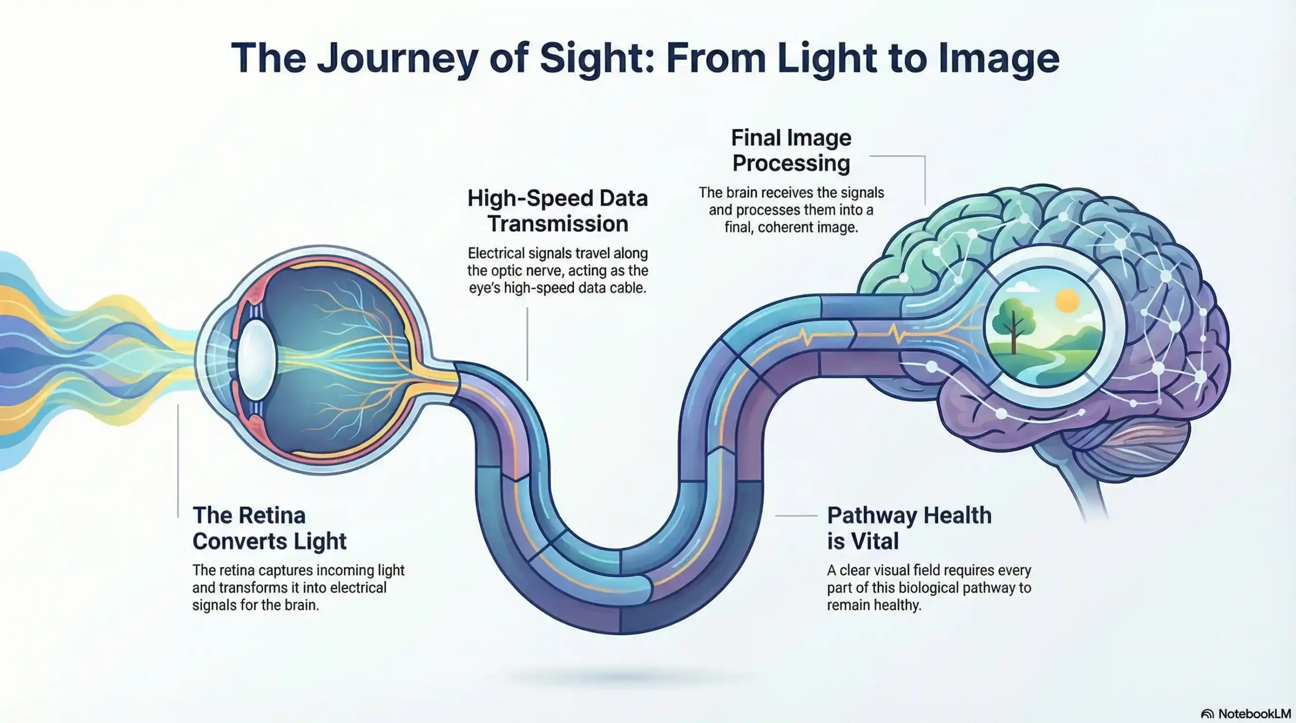
Vision is a complex relay race between your eyes and your brain. When light enters the eye, it strikes the retina, which converts the light into electrical signals. These signals travel along the optic nerve—the “high-speed data cable” of the eye—to the brain, where the image is finally processed.
A clear visual field requires every part of this pathway to be healthy. If the optic nerve is damaged by pressure or if there is a neurological disruption, the “data” from specific parts of your visual field may never reach the brain. This is why testing is so critical; it doesn’t just check the health of your eyes, it monitors the neurological pathways that allow you to see.
Types of Visual Field Vision
Your vision is generally categorized into two main functions:
-
Central Vision: This is the high-resolution vision used for detailed tasks like reading fine print or recognizing faces. It is powered by the macula, the center of your retina.
-
Peripheral (Side) Vision: This occurs outside your direct line of sight. It is highly sensitive to movement and light, helping you maintain balance and spatial awareness.
During a consultation at our Richmond office, Dr. Zaver explains that while central vision is what you notice most, peripheral vision is what keeps you safe. Our testing assesses both, ensuring your “peripheral awareness” is functioning at 100%.
Causes of Visual Field Loss
Visual field loss can stem from various health issues, ranging from localized eye diseases to systemic conditions:
-
Glaucoma: Known as the “silent thief of sight,” it gradually destroys the optic nerve, usually starting with the side vision.
-
Neurological Issues: Strokes, brain tumors, or Multiple Sclerosis (MS) can cause specific patterns of vision loss.
-
Retinal Conditions: Issues like diabetic retinopathy can create “curtains” or shadows in your vision.
-
Binocular Vision Issues: Sometimes, headaches and eye strain are linked to how the eyes work together, which we address through specialized Neurolens therapy.
How to Protect and Preserve Your Visual Field
Preserving your vision is a proactive journey. Here is how you can protect your sight:
-
Schedule Regular Exams: This is the single most important step. We recommend regular pediatric eye exams for children and annual exams for adults.
-
Manage Systemic Health: Conditions like diabetes directly impact the blood vessels in your eyes.
-
Seek Immediate Care for Changes: If you notice sudden shadows, “tunnel vision,” or flashes of light, contact us for emergency eye care immediately.
-
Protect Against UV: Long-term exposure to the sun can damage the retina.
The importance of visual field testing
Visual field testing is one of the most effective diagnostic tests in the detection of glaucoma. This is because when patients are affected by glaucoma, it is usually the peripheral vision that is affected by their condition first. However, it can also be used to detect central or peripheral retinal diseases, eyelid conditions such as drooping, optic nerve damage, and conditions that affect the visual pathways from the optic nerve to the area of the brain where this information is processed into vision.
Visual field testing is also an important part of monitoring for people who are considered to be at risk for vision loss from disease and other problems, including those who have been diagnosed with the following:
- Multiple sclerosis
- Hyperthyroidism
- Pituitary gland disorders
- Central nervous system problems (such as a tumor that may be pressing on the brain)
- Stroke
- Diabetes
- High blood pressure
What to expect from visual field testing
There are a variety of methods that can be used to perform visual field testing, including:
Static automated perimetry. This is where a machine is used to quantify how well the patient is able to detect flashing lights of varying size and brightness in different areas of their visual field, while they concentrate on a central point. The patient responds by pushing a button when they see the light.
Kinetic perimetry. This involves points of light that are fixed in size and intensity and are presented along the patient’s peripheral vision, before being gradually moved inwards to determine their field of vision.
Visual field testing is non-invasive, painless, and doesn’t require patients to have their eyes dilated. The results, which are usually presented in a series of charts, are digital and sent directly to your eye doctor for interpretation. Depending on the outcome of your results, you may be recommended for further diagnostic testing, which could include blood tests. If you have been diagnosed with glaucoma, you will probably be recommended to have several visual field tests each year, which will help your eye doctor monitor the progression of your condition and recommend treatments to slow it.
Citations and Research Resources
This article is based on current medical literature and professional guidelines from leading healthcare organizations. The following sources provided key information about visual field testing procedures, clinical applications, and diagnostic importance:
1. Visual Field Testing for Glaucoma – A Practical Guide
National Center for Biotechnology Information (NCBI) – PMC
This peer-reviewed medical resource from the Community Eye Health Journal provides comprehensive clinical guidance on visual field testing methodologies and interpretation. The article covers the technical aspects of static and kinetic perimetry, reliability factors in testing, and the critical role of visual field assessment in detecting glaucomatous damage. It emphasizes that glaucomatous visual field loss typically isn’t evident until at least 30% of retinal ganglion cell axons have been lost, highlighting the importance of early and regular testing for at-risk patients.
2. Breaking Down Visual Fields in Glaucoma
Review of Optometry – Clinical Practice Guidelines
Published by Review of Optometry, this clinical article discusses current standards in visual field testing, including the evolution from traditional testing methods to modern SITA (Swedish Interactive Thresholding Algorithm) protocols. The resource provides valuable insights into test interpretation, reliability assessment, and the importance of frequent testing for detecting glaucoma progression. It also covers emerging technologies like home-based visual field testing and their potential impact on patient care and disease management.
At Frame & Focus Eye Care, we are committed to helping our neighbors in Richmond, Pecan Grove, and Sugar Land see the full picture of their health.
Ready to prioritize your vision? Schedule an appointment with Dr. Sarah Zaver today.
FAQs
-
This non-invasive test measures your full range of vision, including central and peripheral (side) sight. It helps eye doctors detect blind spots or vision loss caused by conditions like glaucoma or stroke.


